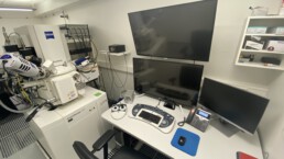
2D and 3D Electron Microscopy, Focused Ion Beam-Scanning Electron Microscopy (Zeiss Crossbeam 340) including Energy dispersive X-ray microscopy (Oxford Instruments)
This electron microscope is suitable for classical secondary electron imaging and backscattered imaging of biological tissues and materials. Additionally, the FIB/SEM mode allows for high resolution volume electron microscopy (~7 nm voxel size) as well as ultrathin lamellae preparation for transmission imaging. Equipped with a STEM detector, lower resolution transmission imaging can be performed within the system at an acceleration voltage of up to 30kV. The system is equipped with an EDX detector for analyzing the elemental composition in biomaterials.
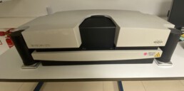
Micro-Computed Tomography (Bruker Skyscan 1727, Scanco µCT-40)
MicroCT imaging is the ideal lab-based imaging tool for analyzing 3D structures at high resolutions. These systems are commonly used to generate morphological information of complex structures such as bone specimens, zebrafish organs, teeth, or other biomaterials at voxel sizes between 0.75 µm and 20 µm. Soft tissue can also be scanned using prior contrast enhancement of specimens.
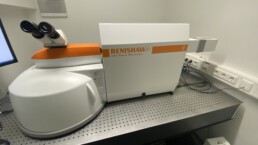
Raman Microspectroscopy Imaging (Renishaw invia)
Raman micro-spectroscopy allows to assess the macromolecular composition of materials and tissues. It is specifically valuable for analyzing mineralized hard tissues as it provides the ability to assess both protein- and mineral-related components of the tissues simultaneously. The system is equipped with a 738 nm red laser, 5X, 20X, and 50X objectives as well as a 60X immersion objective for scanning moist or fresh tissue specimens.
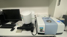
Fourier Transform Infrared spectroscopy (FTIR) imaging (Perkin Elmer Spotlight 400)
Similar to Raman spectroscopy, FTIR is used to assess the macromolecular composition of materials and tissues.
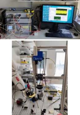
Optical Coherence Tomography (OMES 4D MHz-OCT System)
The OMES OCT system operates at a wavelength of 1315nm and allows us to acquire up to 833 full volumes per second. This high temporal frequency is beneficial in precise tracking of soft-tissue as well as in elasticity estimation by tracking the movement of shear waves inside soft-tissue.
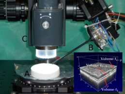
Optical Coherence Tomography (Thorlabs Telesto OCT System)
The Telesto OCT system is an OCT system which operates at wavelength of 1300 nm. A high resolution scan head allows the acquisition of volumes with a size of up to 15x15x3.5mm. Further, optical fibers integrated inside needle can be connected to the system to enable among other things imaging of soft tissue at the needle tip.

Ultrasound Imaging (Cephasonics Ultrasound Imaging System)
The Cephasonics ultrasound platform boasts an impressive 40MHz sampling rate, enabling the high-resolution capture of dynamic processes within the body. With a sophisticated design featuring 256 individual channels, coupled with our custom volumetric ultrasound transducer equipped with a 16x16 element array, we seamlessly acquire 4D data without the necessity for multiplexing and the associated delays in volume acquisition.