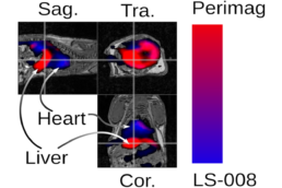
High Resolution Imaging for Skeletal Aging
We research bone fragility and aging with high resolution imaging modalities to enhance patient morbidity and diagnostics.
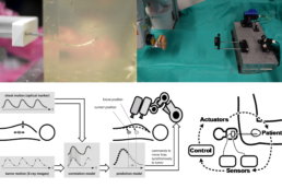
Tissue Interface Sensing during Needle Placement
We research if tissue interfaces can be sensed by the surgeon while purely relying on haptic feedback through a robot. Thereby needle placement relative to known tissue interfaces is improved. We sense forces at the needle tip with our in house build needle probes and perform real-time data processing with neural networks. The sensed forces can be felt by the physician at the robotic handle. The system was tested in-situ with medical experts.
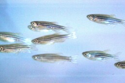
Motion Analysis for monitoring musculoskeletal health in a Fish Model
Similar to humans, the physical mobility of zebrafish can be impaired due to aging or disease (e.g. osteoarthritis, scoliosis). While pathological locomotion patterns of individual fish may appear obvious to the observer, visual inspection remains difficult because of multiple moving objects, rendering the quantification of changes in mobility impossible. We design machine learning tracking algorithms to study the movement patterns of fish over long time periods and evaluate the physical performance, locomotion phenotypes, and treatment strategies in zebrafish models of skeletal disease.
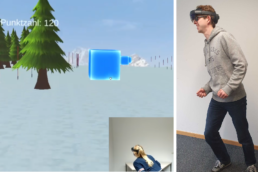
Virtual reality-based Patient Training
Prevention of falls accompanied by severe injuries such as fracture is of high importance for our aging society. Beyond muscle function, bone quantity and quality-indices, in particular body-control and -coordination are considered to have major impact on preventing fractures by minimizing fall-events themselves. We investigate how virtual reality applications can enhance the training of elderly and reduce bone fractures.
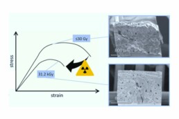
Influence of X-ray radiation on tissues
Doses of irradiation above 25 kGy are known to cause irreversible mechanical decay in bone tissue. However, the impact of irradiation doses absorbed in a clinical setting on the mechanical properties of bone remains unclear. In daily clinical practice and research, patients and specimens are exposed to irradiation due to diagnostic imaging tools, with doses ranging from milligray to Gray. The aim of this study was to investigate the influence of irradiation at these doses ranges on the mechanical performance of bone independent of inter-individual bone quality indices.
Machine Learning
We study and apply various machine learning methods (e.g., CBR,SVM/SVR) in a number of clinical contexts, with one focus on time series analysis. Moreover we design deep and convolutional neural networks for classification and regression tasks based on high-frequency sensor and clincial image data. Particularly, we extend deep learning methods to spatio-temporal data, including 4D image data. Application examples range from nephrology to tumor tissue detection to cardiology.
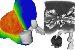
Robotics
We study methods to control and navigate robot motion, particularly for medical applications. Examples include robotic radiosurgery, robotic transcranial stimulation and robotic needle placement. We are interested in approaches to detect and compensate motion and deformation, e.g., due to respiration, pulsation, or soft tissue deformation. Moreoever, we are also interested in human-robot interaction and mobile robotics. At our lab, we have a number of navigation systems (e.g., optical and electro-magnetic) and various robots (starting from 0.01 mm repeatability) to realize experimental Setups.
Navigation
With our high-frequency ultrasound, optical coherence and optical tracking systems we can systematically analyze motion and compensate for displacements through real-time data processing. Predicting movements and compensating them with a robot is one of our research areas, e.g. for steering a needle in soft tissue.
Planning and Optimization
We apply planning and optimization approaches in several research areas, including treatment planning for radiation therapy and brachytherapy, as well as optimization of robot configurations and movements.
Radiation therapy presents an effective and non-invasive option for cancer treatment. However, ionizing radiation can be harmful and the effects and side effects of irradiation need to be balanced. We also examine robotic biopsy, which similarly requires careful planning for safe and time efficient tissue sampling.
We develop algorithms and systems involving heuristic optimization, mathematical programming, and machine learning to identify the optimal trade-off among the various clinical objectives.

Multi-scale Analysis of Tissue Hierarchy
Bone is a natural, hierarchical composite material consisting of proteins and minerals (mainly mineralized collagen fibrils), and water. Mineralized tissues including bones and teeth gain their high fracture resistance through a tissue-specific hierarchical makeup, and changes at any length scale can have an influence on the mechanical properties at the skeletal level. Analyzing the structure, composition, and mechanical properties of bone tissue helps to better understand bone in healthy and diseased conditions.
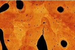
Imaging of Musculoskeletal Tissues
Advanced 2D and 3D imaging techniques allow the visualization of tissue changes at multiple length scales. By combining imaging tools such as high-resolution peripheral quantitative computed tomography (HR-pQCT), microCT, and electron microscopy (FIB/SEM) we can visualize various features of bone tissue including cortical and trabecular components, vascular or lacunar porosities, cell organelles, collagen fibrils and mineral particles.
Locomotion patterns and skeletal function
Over the course of evolution, different types of locomotion have emerged within vertebrate species, for example, swimming in bony fish, quadrupedal walking in mice, and bipedal walking in humans. Despite these differences, the fundamental composition of bone tissue as well as its genetic regulation has been largely conserved across species. We can detect specific structure-function relationships of mineralized tissues across species to better understand the musculoskeletal system.

Reconstruction-Based Artifact Reduction
In imaging, a variety of factors, such as technical imperfections, the complexity of the underlying physics or patient motion, can be a cause of artifacts. In many cases, the latter can be mitigated by a suitable extension of the established standard image reconstruction algorithms. Recently, we also started exploring modern data-driven approaches for the mitigation of artifacts.
Multi-Contrast Imaging
While standard imaging often generates images with a single contrast, modern acquisition schemes also allow to obtain multiple image contrasts from a single acquisition. Importantly, this can lead to an increased diagonistic value by opening up new ways of characterizing the object being imaged. An important aspect of our research is the development and improvement of the image reconstruction methods required to enable multi-contrast imaging.
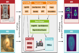
Image Reconstruction Software
Modern imaging relies on more and more sophisticated image reconstruction algorithms, which often have a high computational complexity. To facilitate the development and the adoption of new image reconstruction methods, we develop suitable image reconstruction frameworks, which are both performant and highly accessible.