Reserach @ MTEC
In our research projects we develop, extend and adapt methods from robotics and navigation, search and optimization, machine learning, and signal and image processing to realize systems and system components for clinical applications ranging from tissue characterization to radiosurgical treatments.
 Motion Prediction and Compensation
With our high-frequency ultrasound, optical coherence and optical tracking systems we can systematically analyze motion and compensate for displacements through real-time data processing. Predicting movements and compensating them with a robot is one of our research areas, e.g. for continuous compensation of head movements during transcranial magnetic stimulation.
Motion Prediction and Compensation
With our high-frequency ultrasound, optical coherence and optical tracking systems we can systematically analyze motion and compensate for displacements through real-time data processing. Predicting movements and compensating them with a robot is one of our research areas, e.g. for continuous compensation of head movements during transcranial magnetic stimulation.
![]()
Motion Prediction and Compensation
With our high-frequency ultrasound, optical coherence and optical tracking systems we can systematically analyze motion and compensate for displacements through real-time data processing. Predicting movements and compensating them with a robot is one of our research areas, e.g. for continuous compensation of head movements during transcranial magnetic stimulation.
Collaborative Robotics
We develop robotic systems that combine sensing with real-time data processing through machine learning to enable precise positioning of a medical tool with a robot. Customized sensors and haptic feedback enable the user to perceive new or enhanced sensations, allowing the surgeon to feel, for example, tissue transitions during collaborative procedures. Commonly, we test our systems in our laboratory and with clinical physicians.
![]()
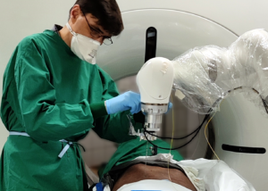
Motion Prediction and Compensation
With our high-frequency ultrasound, optical coherence and optical tracking systems we can systematically analyze motion and compensate for displacements through real-time data processing. Predicting movements and compensating them with a robot is one of our research areas, e.g. for continuous compensation of head movements during transcranial magnetic stimulation.
![]()
Motion Prediction and Compensation
With our high-frequency ultrasound, optical coherence and optical tracking systems we can systematically analyze motion and compensate for displacements through real-time data processing. Predicting movements and compensating them with a robot is one of our research areas, e.g. for continuous compensation of head movements during transcranial magnetic stimulation.
Reserach @ Heisenberg Research Group
The fracture risk of bone is not solely dependent on bone quantity; indeed, bone’s hierarchical structure has distinct features at multiple length-scales, and thus the quality of the structure plays a large role in its resistance to fracture. Understanding how the nature of fracture is affected by bone quality…
 Analysis of Bone Material Quality
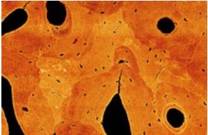
Bone is a natural, hierarchical composite material consisting of proteins and minerals (mainly mineralized collagen fibrils), and water. Mineralized tissues including bones and teeth gain their high fracture resistance through a tissue-specific hierarchical makeup, and changes at any length scale can have an influence on the mechanical properties at the skeletal level. Analyzing the structure, composition, and mechanical properties of bone tissue helps to better understand bone in healthy and diseased conditions.
Multi-scale Imaging of Musculoskeletal Tissues
Advanced 2D and 3D imaging techniques allow the visualization of tissue changes at multiple length scales. By combining imaging tools such as high-resolution peripheral quantitative computed tomography (HR-pQCT), microCT, and electron microscopy (FIB/SEM) we can visualize various features of bone tissue including cortical and trabecular components, vascular or lacunar porosities, cell organelles, collagen fibrils and mineral particles.
Locomotion patterns and skeletal function
Over the course of evolution, different types of locomotion have emerged within vertebrate species, for example, swimming in bony fish, quadrupedal walking in mice, and bipedal walking in humans. Despite these differences, the fundamental composition of bone tissue as well as its genetic regulation has been largely conserved across species. We can detect specific structure-function relationships of mineralized tissues across species to better understand the musculoskeletal system.
Research @ IBI
Research projects at the IBI cover imaging modalities such as magnetic particle imaging, magnetic resonance imaging and computed tomography. A special focus is placed on the development of image reconstruction techniques, which allow for an acceleration of measurements, mitigation of artifacts or the generation of new imaging contrasts.
Reconstruction-Based Artifact Reduction
In imaging, a variety of factors, such as technical imperfections, the complexity of the underlying physics or patient motion, can be a cause of artifacts. In many cases, the latter can be mitigated by a suitable extension of the established standard image reconstruction algorithms. Recently, we also started exploring modern data-driven approaches for the mitigation of artifacts.
Multi-Contrast Imaging
While standard imaging often generates images with a single contrast, modern acquisition schemes also allow to obtain multiple image contrasts from a single acquisition. Importantly, this can lead to an increased diagonistic value by opening up new ways of characterizing the object being imaged. An important aspect of our research is the development and improvement of the image reconstruction methods required to enable multi-contrast imaging.
Image Reconstruction Software
Modern imaging relies on more and more sophisticated image reconstruction algorithms, which often have a high computational complexity. To facilitate the development and the adoption of new image reconstruction methods, we develop suitable image reconstruction frameworks, which are both performant and highly accessible.
Reserach @ Institut für Biomedizinische Bildgebung
The Institute of Biomedical Imaging is a joint research department between the University Medical Center Hamburg-Eppendorf (UKE) and the Hamburg University of Technology. The research interests are in signal processing of tomographic imaging techniques. In particular image reconstruction and image computing are the key competences of our research group. In close collaboration with our clinical partners at the UKE we further do translational research with the goal of bringing state of the art acquisition and reconstruction methods into the clinical routine.
Project
…
Project
…
Project
…
Project
…
Project
…
Project
…